
The brain regulates every critical bodily function, from movement and digestion to thought and respiration. But how does the brain, which sits at the very top of your body in your skull, command everything from your eyes to your heart to your feet?
The brain communicates with and controls every part of the human body through a network of neurons called the peripheral nervous system. This network runs across the whole body, communicating the signals between the different organs to and from the brain.
The neuron is the primary functional unit of the peripheral nervous system responsible for relaying signals from all parts of the body to the brain, where they are processed and sent back to their source.
Let’s look at the neuron’s structure and how it communicates with other neurons.
Anatomy Of The Neuron
The structure of the neuron allows for efficient information transmission and reception throughout the entire organism. The Spanish neuroscientist Santiago Ramón y Cajal, also known as the “father of neuroscience,” is widely credited for discovering the structure and many other properties of neurons.
In 1906, Ramón y Cajal became the first Spaniard to win the Nobel Prize in Physiology or Medicine for his work on neurons and the development of the neuron doctrine, which is considered to be the cornerstone of modern neuroscience.
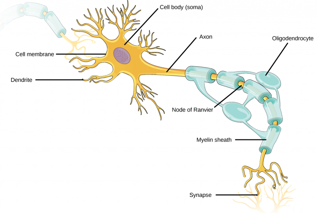
The structure comprises three primary components: the cell body, axon, and dendrites. Let’s learn about each part and its significance in communication.
Cell Body
The nucleus, located in the cell body (sometimes called the “soma”), is the neuron’s central hub for protein production.
Dendrites
Dendrites are protruding branches from the cell body that receive signals from other neurons via the axon terminal.
Axon
An axon is an extended part of a cell that conducts action potentials and other nerve impulses away from the cell body along its cable-like structure. The end of the axon, called the axon terminal, is indirectly attached to the adjacent neuron’s dendrite via a synapse that plays a crucial part in neuron communication.
The axons are insulated with a fatty layer of the myelin sheath, like insulation on a wire. Along the length of a myelinated axon, there are bare spots known as nodes of Ranvier. Axonal saltatory conduction is facilitated by the myelin sheath and the nodes of Ranvier, allowing for rapid transmission of electrical impulses along the axon. In this form of conduction, the action potential skips over the axonal nodes of Ranvier en route to the terminal, shortening the time required for the impulse to travel the whole length of the axon.
Damage to this myelinated sheath disrupts the transmission of action potentials, leading to multiple sclerosis.
Apart from neurons, the peripheral nervous system also comprises glial cells that support neurons in transmitting and receiving electrical and chemical impulses. They maintain homeostasis, produce myelin in the peripheral nervous system, feed neurons with fuel and oxygen, and shield and sustain the cells. Oligodendrocytes are glial cells that aid in the development of myelin for the neurons they are linked to, as shown in the figure above.
Communication Between Neurons
Connectivity between neurons is essential for the execution of all of our conscious and unconscious behaviors. The neurons are able to communicate with one another via both electrical and chemical signals. Let us know more about the two types of transmission.
Electrical Transmission
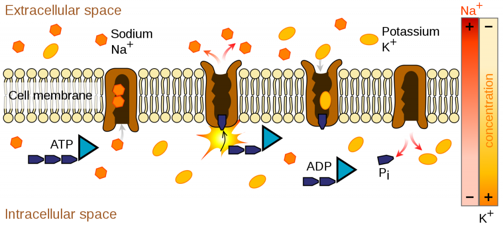
A neuron, like every other cell in our bodies, has its own selectively permeable lipid-bilayer membrane. There are ion channels in the membrane that let electrically charged ions pass through, and ion pumps that chemically move ions from one side of the membrane to the other. Ions such as Na+, K+, Cl-, and Ca2+ constantly flow to and from the ion channel within and outside of the neuron in response to neuronal activity. A membrane potential, or action potential, is generated as a result of this disparity in ion concentration between the interior and exterior of the neuron.
When the neuron is at rest, it seeks to maintain its resting potential by allowing ions to flow via its ion channels. A sodium-potassium pump located in the neuron membrane expels sodium ions and imports potassium ions. However, there is a tiny negative charge within the cell because the number of sodium ions leaving the cell is greater than the number of potassium ions entering the cell. There are also sodium and potassium leak channels that allow the diffusion of these ions across the membrane, but because there are more potassium leak channels than sodium leak channels, more positively charged ions are leaving the neuron than entering, resulting in a net negative charge on the neuron. This ion flow is responsible for a neuron’s typical resting charge of -70 mV.
When a neuron receives electrical input from another neuron, or “activates,” these ions travel inside the neuron to raise the membrane potential. Neurotransmitters are the chemical messages that neurons receive from other neurons. This leads to the opening of voltage-gated ion channels, resulting in membrane potential being more negative or more positive depending on the ions that flow in. When a neurotransmitter triggers the opening of voltage-gated sodium channels, sodium ions flow into the neuron, elevating the membrane potential, while when a potassium channel is activated, potassium ions flow out, depressing the membrane potential.
The following steps must take place in order for information to be sent effectively from one neuron to the next:
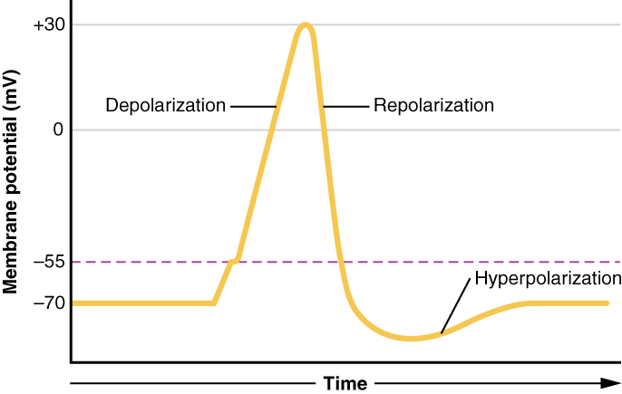
- Depolarization: As the name suggests, depolarization occurs when the membrane potential rises due to an inflow of sodium ions when voltage-gated sodium channels are opened, although not every depolarization results in a conductive action potential. If a neuron is to enter its active state, its membrane potential must rise above the threshold potential (about -55 mV). A successful depolarization can make the membrane potential go as high as 30 mV.
- Repolarization: When the action potential has peaked, the sodium channels close, and the potassium channels open, with a delay of a few milliseconds in between. This process is called repolarization. As a result, potassium ions diffuse out of the cell, creating a negative membrane potential.
- Hyperpolarization: As the membrane potential falls below the resting potential, hyperpolarization occurs and potassium ions continue to rush out. This is called hyperpolarization. Sodium channels open at this point to restore the membrane potential to its resting value. The neuron is ready to fire again because the sodium-potassium pump and leak channels have opened to keep the ion concentration the same as in the dormant state.
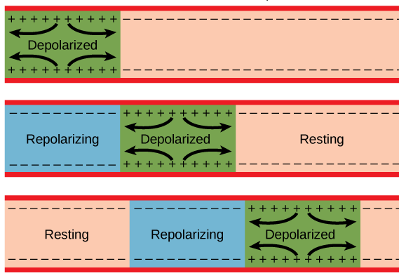
These stages of polarization occur along the length of the axon, depolarizing each node along the way.
Chemical Transmission
Synapses are junctions between the dendrites of one neuron and the axon terminals of neighboring neurons. It is the presynaptic neuron that transmits the signal to the next cell and the postsynaptic neuron that receives the signal. The synaptic cleft is the space between a presynaptic neuron and a postsynaptic neuron, and it is here that neurotransmitters are released. Neurotransmitters are signaling molecules in the axon terminal that carry signals to other neurons.
Depolarization of the terminal membrane caused by the action potential traveling through the axon allows sodium ions to diffuse through the membrane. Depolarization triggers the opening of calcium channels present at the terminal’s periphery. Synaptic vesicles, which contain numerous neurotransmitters, fuse with the terminal membrane in response to the influx of calcium ions. As a result, neurotransmitters are released into the synaptic cleft, signaling the postsynaptic neuron of the ensuing communication stream.
Once released, neurotransmitters bind to receptor proteins on the dendrites of postsynaptic neurons. Receptors are protein-based chemical structures that act on the cells they are present on in response to incoming signals. Whether a postsynaptic cell becomes depolarized or hyperpolarized depends on the nature of the neurotransmitter and its receptor. When a neurotransmitter causes sodium channels to open, the neuron depolarizes, producing an EPSP (Excitatory PostSynaptic Potential) that increases the likelihood that the neuron will fire an action potential. If, however, it causes chlorine channels to open, the neuron will hyperpolarize, resulting in an IPSP (Inhibitory Postsynaptic Potential) that reduces the likelihood of action potential firing. A neuron’s decision to send an action potential down its axon is based on the sum of the inhibitory and excitatory potentials it receives from all of its dendritic spines.
The human nervous system contains over a hundred different types of neurotransmitters.
Once the signal has been transmitted to the postsynaptic neuron, the neurotransmitters must be removed from the cleft to return the synapse to its resting state and prevent further depolarization of the postsynaptic neuron. There are three ways elimination of these molecules occurs:
- Diffusion: They are destroyed by the glial cells after diffusing out of the synaptic cleft.
- Enzyme Degradation: Enzymes present in the cleft can break down the neurotransmitters in the cleft itself.
- Reuptake: They can be reabsorbed by the presynaptic neuron for reuse in another firing event.
When brain activity is recorded, a single cycle through the aforementioned electrical and chemical transmission results in a spike that may be used to decode which neurons were engaged during a given task. This has numerous implications in the scientific and technological fields, leading to devices that might aid persons suffering from ailments caused by disturbances in communication patterns.
Follow The Brainy Bits for more interesting content